|
|
|
|
|
|

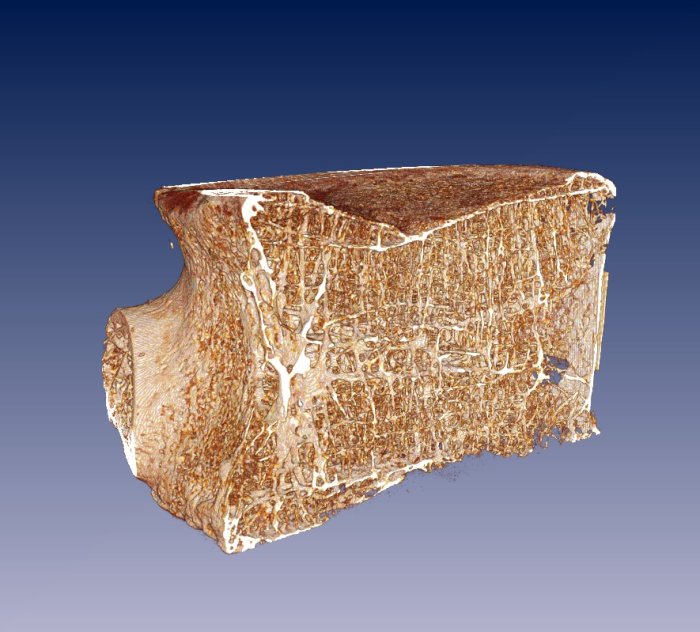
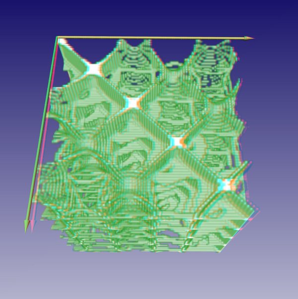
Changes in trabecular bone composition during the development of osteoporosis are used as a model of bone loss in microgravity. The structural changes of trabecular bone have received more attention in the last years as bone densitometry alone cannot explain all variation in bone strength. We have previously successfully developed and applied a set of measures of complexity, which quantify the trabecular bone architecture using 2D Computed Tomography (CT) images. The rapid progress in high resolution 3D microCT imaging facilitates the development of new 3D measures of complexity, which should be able to assess the spatial architecture of trabecular bone. Currently, we follow different approaches, based on spatial correlation, translational invariance, recurrence of spatial structures, curvatures and surface geometry.
Further reading:
Gallery: